Exhibit 99.1
MelaFind®
Optics by Carl Zeiss
Needham Growth Conference
MELA Sciences, Inc.
January 16, 2013
©MELA Sciences 2012
Forward Looking Statements
This presentation includes “forward-looking statements” within the meaning of the Securities Litigation Reform Act of 1995. These statements include but are not limited to our plans, objectives, expectations and intentions and other statements that contain words such as “expects,” “contemplates,” “anticipates,” “plans,” “intends,” “believes” and variations of such words or similar expressions that predict or indicate future events or trends, or that do not relate to historical matters. These statements are based on our current beliefs or expectations and are inherently subject to significant uncertainties and changes in circumstances, many of which are beyond our control. There can be no assurance that our beliefs or expectations will be achieved. Actual results may differ materially from our beliefs or expectations due to economic, business, competitive, market and regulatory factors. The proposed offering is confidential. Anyone receiving or viewing the Management Presentation should keep it confidential and not use it for any purpose other than to consider an investment in the Company in connection with the offering.
MELA: Nasdaq CM
©MELA Sciences 2012
MelaFind®
Optics by Carl Zeiss
2
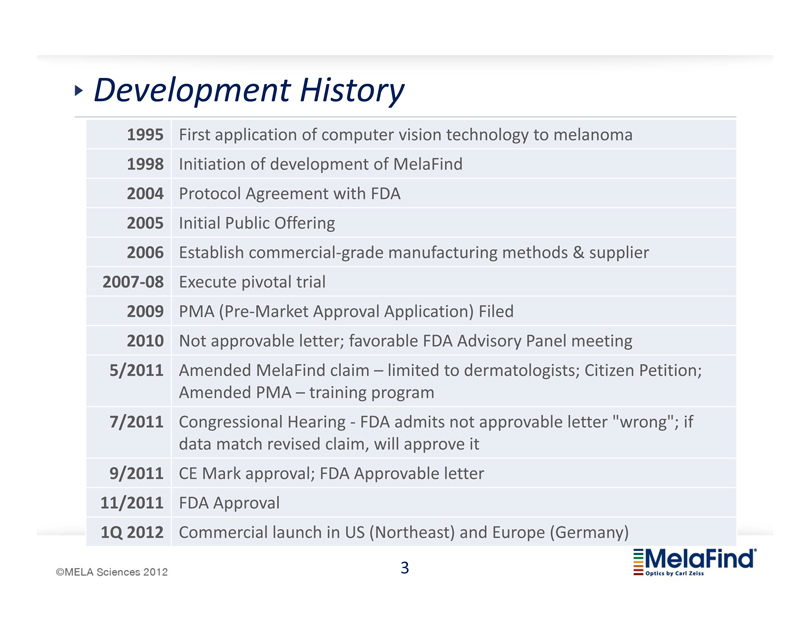
Development History
1995 First application of computer vision technology to melanoma
1998 Initiation of development of MelaFind
2004 Protocol Agreement with FDA
2005 Initial Public Offering
2006 Establish commercial-grade manufacturing methods & supplier
2007-08 Execute pivotal trial
2009 PMA (Pre-Market Approval Application) Filed
2010 Not approvable letter; favorable FDA Advisory Panel meeting
5/2011 Amended MelaFind claim – limited to dermatologists; Citizen Petition;
Amended PMA – training program
7/2011 Congressional Hearing - FDA admits not approvable letter “wrong”; if
data match revised claim, will approve it
9/2011 CE Mark approval; FDA Approvable letter
11/2011 FDA Approval
1Q 2012 Commercial launch in US (Northeast) and Europe (Germany)
©MELA Sciences 2012
MelaFind®
Optics by Carl Zeiss
3
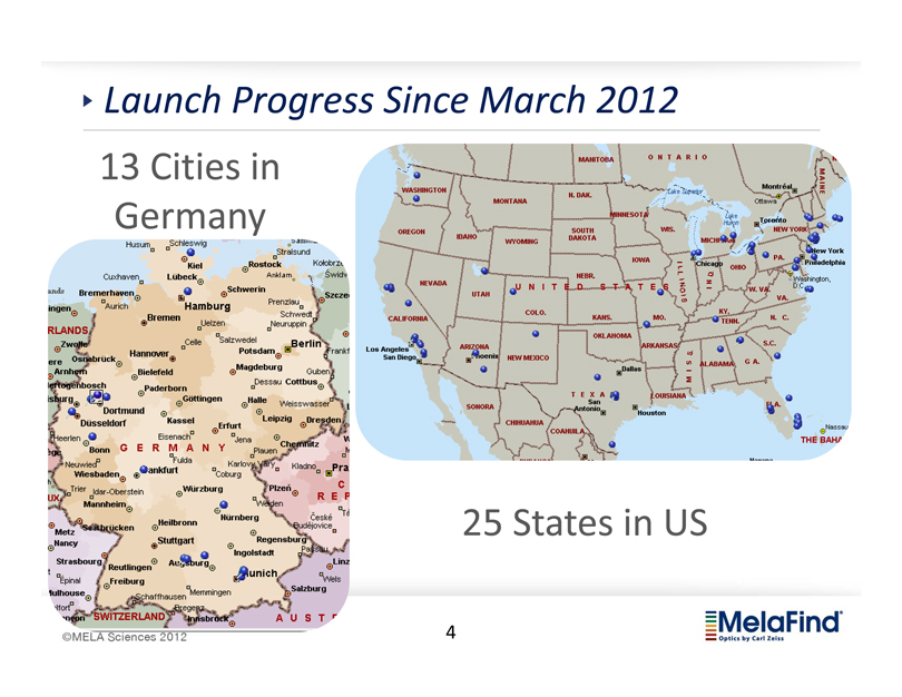
Launch Progress Since March 2012
13 Cities in Germany
25 States in US
©MELA Sciences 2012
MelaFind®
Optics by Carl Zeiss
4
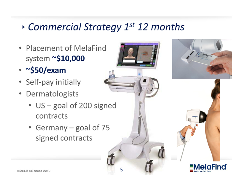
Commercial Strategy 1st 12 months
• Placement of MelaFind system ~$10,000
• ~$50/exam
• Self-pay initially
• Dermatologists
• US – goal of 200 signed contracts
• Germany – goal of 75 signed contracts
©MELA Sciences 2012
MelaFind®
Optics by Carl Zeiss
5
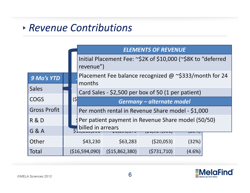
Revenue Contributions
9 Mo’s YTD
Sales
COGS
Gross Profit
R & D
G & A
Other
Total
ELEMENTS OF REVENUE
Initial Placement Fee:
$2K of $10,000 (
$8K to “deferred revenue”)
Placement Fee balance recognized @
$333/month for 24 months
Card Sales - $2,500 per box of 50 (1 per patient)
($ Germany – alternate model
Per month rental in Revenue Share model - $1,000
Per patient payment in Revenue Share model (50/50) ($ billed in arrears
$43,230
$63,283
($20,053)
(32%)
($16,594,090)
($15,862,380)
($731,710)
(4.6%)
©MELA Sciences 2012
MelaFind®
Optics by Carl Zeiss
6
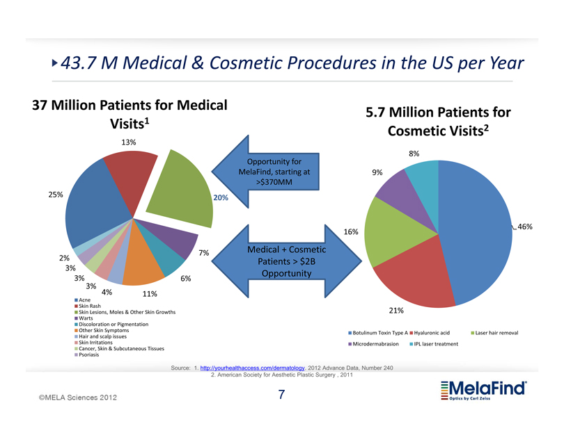
43.7 M Medical & Cosmetic Procedures in the US per Year
37 Million Patients for Medical Visits1
13%
25% 20%
7%
2%
3%
3% 6%
3% 4% 11%
Acne
Skin Rash
Skin Lesions, Moles & Other Skin Growths
Warts
Discoloration or Pigmentation
Other Skin Symptoms
Hair and scalp issues
Skin Irritations
Cancer, Skin & Subcutaneous Tissues
Psoriasis
Opportunity for MelaFind, starting at >$370MM
Medical + Cosmetic Patients > $2B Opportunity
5.7 Million Patients for Cosmetic Visits2
8%
9%
46%
16%
21%
Botulinum Toxin Type A
Hyaluronic acid
Laser hair removal
Microdermabrasion
IPL laser treatment
Source: 1. http://yourhealthaccess.com/dermatology. 2012 Advance Data, Number 240
2. American Society for Aesthetic Plastic Surgery , 2011
MELA Sciences 2012
MelaFind
Optics by Carl Zeiss
7
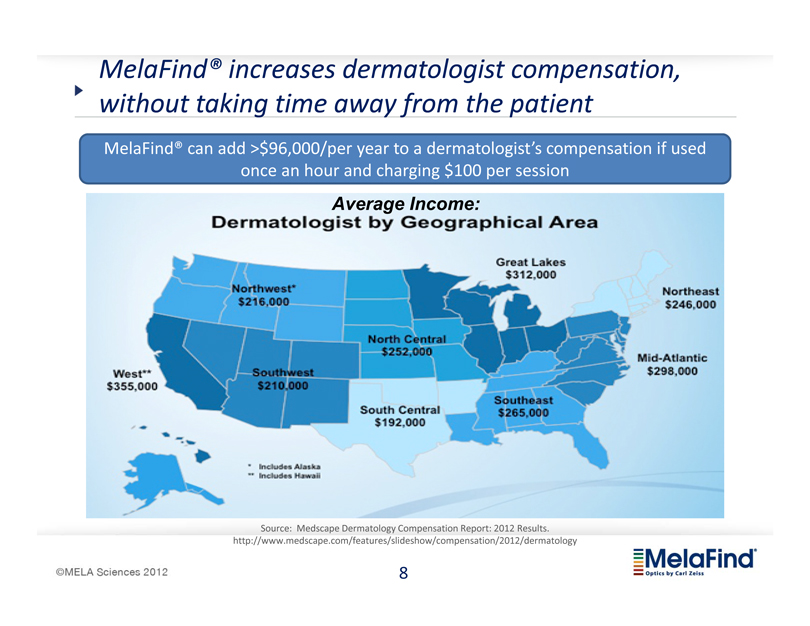
MelaFind increases dermatologist compensation, without taking time away from the patient
MelaFind can add >$96,000/per year to a dermatologist’s compensation if used once an hour and charging $100 per session
Average Income:
Dermatologist by Geographical Area
Source: Medscape Dermatology Compensation Report: 2012 Results.
http://www.medscape.com/features/slideshow/compensation/2012/dermatology
MELA Sciences 2012
MelaFind
Optics by Carl Zeiss
8
2012 Accomplishments
Launched MelaFind (March)
Product upgrades
Developed infrastructure for national business
Many examples of clinical benefit of MelaFind
Melanomas detected
Unnecessary biopsies averted
Ended year with over 100 signed user agreements
Cleveland Clinic Top 10 Innovations for 2013; other unsolicited media attention (print and TV)
German Reader Study results
Initiated FDA post-Approval study
Refined messaging and strategies for increased usage
MELA Sciences 2012
MelaFind
Optics by Carl Zeiss
9
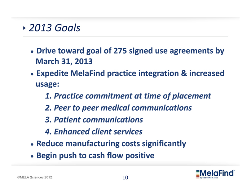
2013 Goals
Drive toward goal of 275 signed use agreements by March 31, 2013
Expedite MelaFind practice integration & increased usage:
1. Practice commitment at time of placement
2. Peer to peer medical communications
3. Patient communications
4. Enhanced client services
Reduce manufacturing costs significantly
Begin push to cash flow positive
MELA Sciences 2012
MelaFind
Optics by Carl Zeiss
10
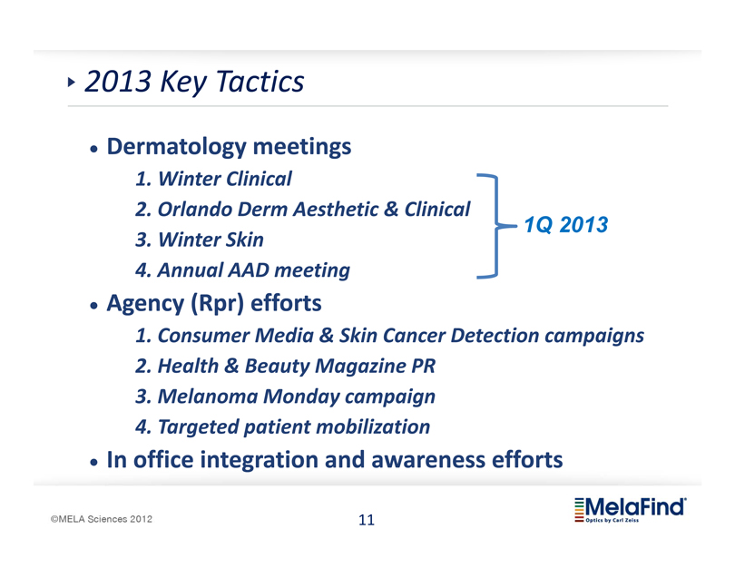
2013 Key Tactics
Dermatology meetings
1. Winter Clinical
2. Orlando Derm Aesthetic & Clinical
1Q 2013
3. Winter Skin
4. Annual AAD meeting
Agency (Rpr) efforts
1. Consumer Media & Skin Cancer Detection campaigns
2. Health & Beauty Magazine PR
3. Melanoma Monday campaign
4. Targeted patient mobilization
In office integration and awareness efforts
MELA Sciences 2012
MelaFind
Optics by Carl Zeiss
11
Rpr Client & Industry Experience
MELA Sciences 2012
MelaFind
Optics by Carl Zeiss
12
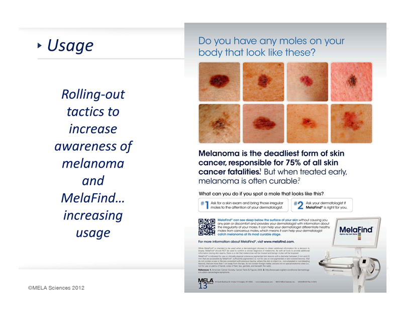
Usage
Rolling-out tactics to increase awareness of melanoma and MelaFind… increasing usage
Do you have any moles on your body that look like these?
Melanoma is the deadliest form of skin cancer, responsible for 75% of all skin cancer fatalities.1 But when treated early, melanoma is often curable.2
MELA Sciences 2012
13
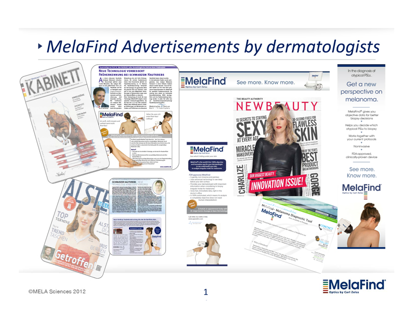
MelaFind Advertisements by dermatologists
MELA Sciences 2012
MelaFind
Optics by Carl Zeiss
1
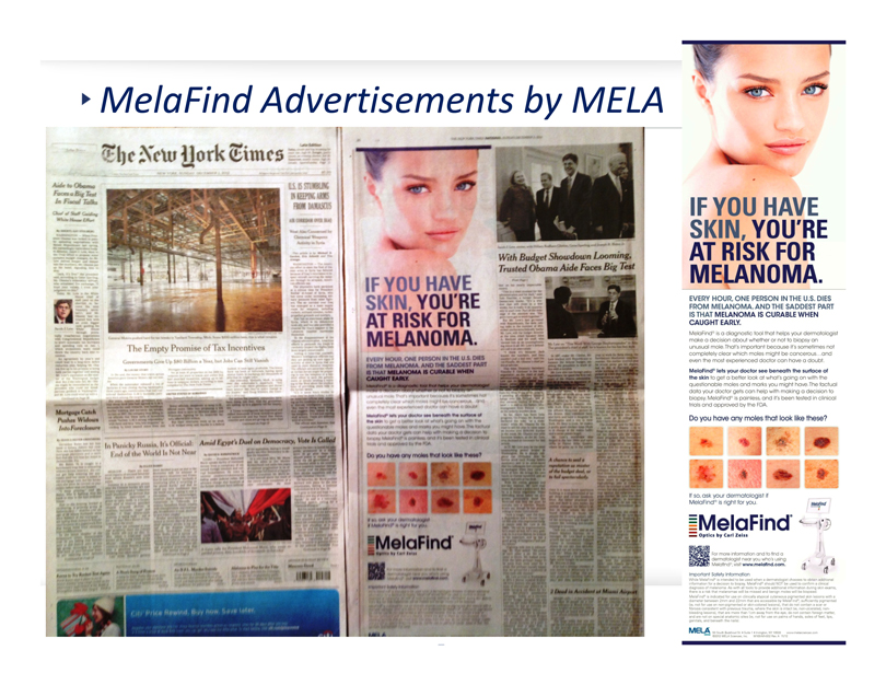
MelaFind Advertisements by MELA
IF YOU HAVE SKIN, YOU’RE AT RISK FOR MELANOMA.
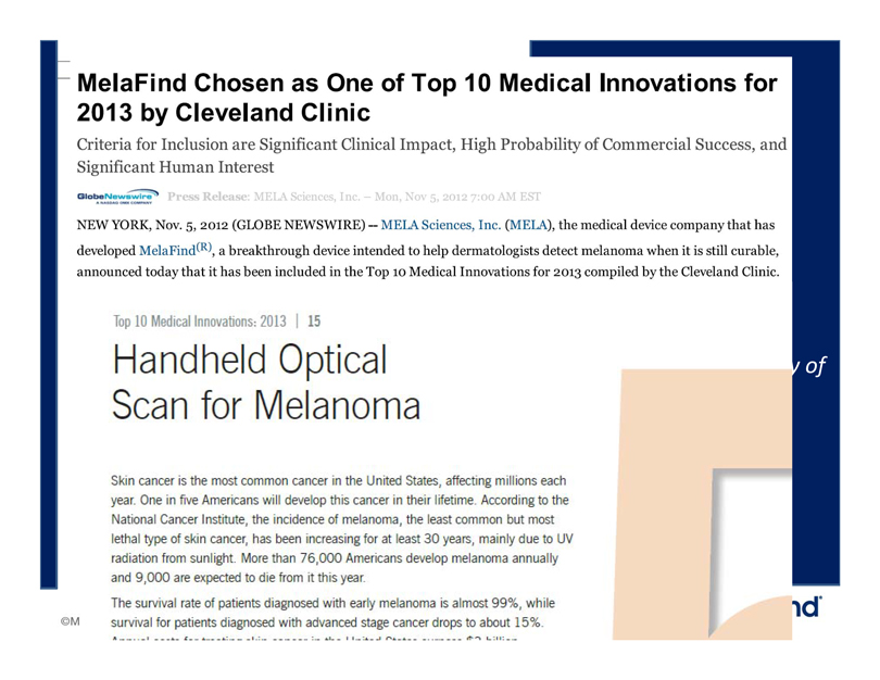
MelaFind Chosen as One of Top 10 Medical Innovations for
2013 by Cleveland Clinic
Criteria for Inclusion are Significant Clinical Impact, High Probability of Commercial Success, and Significant Human Interest
Press Release: MELA Sciences, Inc. – Mon, Nov 5, 2012 7:00 AM EST
NEW YORK, Nov. 5, 2012 (GLOBE NEWSWIRE) — MELA Sciences, Inc. (MELA), the medical device company that has developed MelaFind®, a breakthrough device intended to help dermatologists detect melanoma when it is still curable, announced today that it has been included in the Top 10 Medical Innovations for 2013 compiled by the Cleveland Clinic.
Top 10 Medical Innovations: 2013 | 15
Handheld Optical
Scan for Melanoma
Skin cancer is the most common cancer in the United States, affecting millions each year. One in five Americans will develop this cancer in their lifetime. According to the National Cancer Institute, the incidence of melanoma, the least common but most lethal type of skin cancer, has been increasing for at least 30 years, mainly due to UV radiation from sunlight. More than 76,000 Americans develop melanoma annually and 9,000 are expected to die from it this year.
The survival rate of patients diagnosed with early melanoma is almost 99%, while survival for patients diagnosed with advanced stage cancer drops to about 15%.
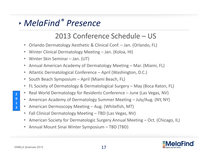
MelaFind ® Presence
2013 Conference Schedule – US
Orlando Dermatology Aesthetic & Clinical Conf. – Jan. (Orlando, FL)
Winter Clinical Dermatology Meeting – Jan. (Koloa, HI)
Winter Skin Seminar – Jan. (UT)
Annual American Academy of Dermatology Meeting – Mar. (Miami, FL)
Atlantic Dermatological Conference – April (Washington, D.C.)
South Beach Symposium – April (Miami Beach, FL)
FL Society of Dermatology & Dermatological Surgery – May (Boca Raton, FL)
Real World Dermatology for Residents Conference – June (Las Vegas, NV)
American Academy of Dermatology Summer Meeting – July/Aug. (NY, NY)
American Dermoscopy Meeting – Aug. (Whitefish, MT)
Fall Clinical Dermatology Meeting – TBD (Las Vegas, NV)
American Society for Dermatologic Surgery Annual Meeting – Oct. (Chicago, IL)
Annual Mount Sinai Winter Symposium – TBD (TBD)
2013
MelaFind® Presence
Upcoming Dermatology Conferences – Germany
Dermatologische Praxis – Mar. (Frankenthal, Germany)
47th DDG-Session – May (Tagung, Germany)
European PostASCO Meeting – TBD (Germany)
Practical Dermatology & Venerology Conference (FOBI) – TBD (Germany)
8th World Congress of Melanoma – July (Hamburg, Germany)
German Skin Cancer Society Meeting (ADO) – Sept (Germany)
2013
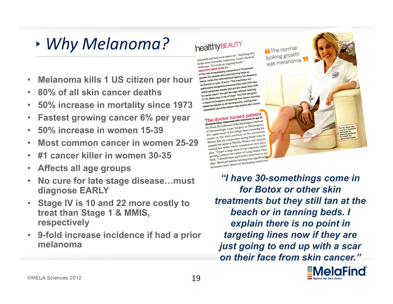
Why Melanoma?
Melanoma kills 1 US citizen per hour
80% of all skin cancer deaths
50% increase in mortality since 1973
Fastest growing cancer 6% per year
50% increase in women 15-39
Most common cancer in women 25-29
#1 cancer killer in women 30-35
Affects all age groups
No cure for late stage disease…must diagnose EARLY
Stage IV is 10 and 22 more costly to treat than Stage 1 & MMIS, respectively
9-fold increase incidence if had a prior melanoma
“I have 30-somethings come in for Botox or other skin treatments but they still tan at the beach or in tanning beds. I explain there is no point in targeting lines now if they are just going to end up with a scar on their face from skin cancer.”
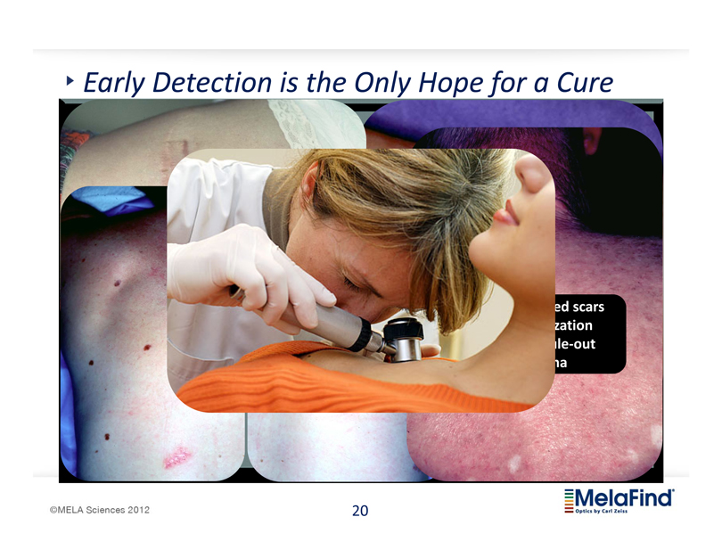
Early Detection is the Only Hope for a Cure
ed scars
zation
ule-out
a
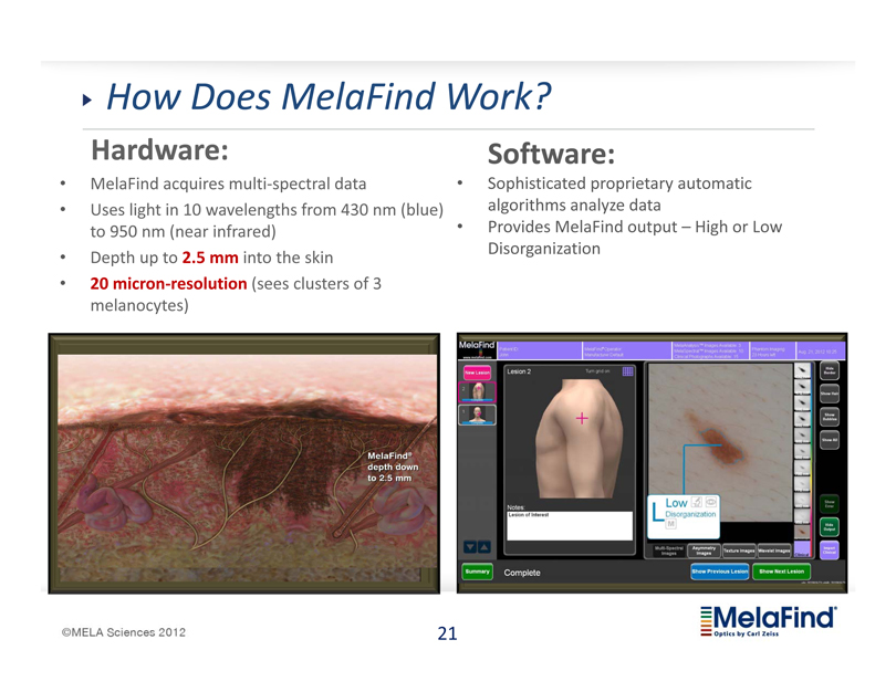
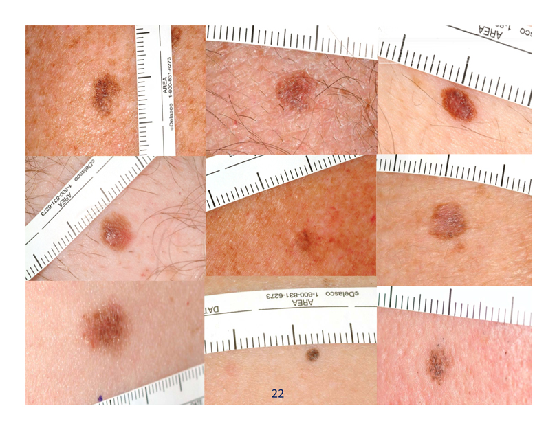
How Does MelaFind Work?
Hardware:
MelaFind acquires multi?spectral data
Uses light in 10 wavelengths from 430 nm (blue) to 950 nm (near infrared) Depth up to 2.5 mm into the skin
20 micron?resolution (sees clusters of 3 melanocytes)
Software:
Sophisticated proprietary automatic algorithms analyze data Provides MelaFind output – High or Low Disorganization
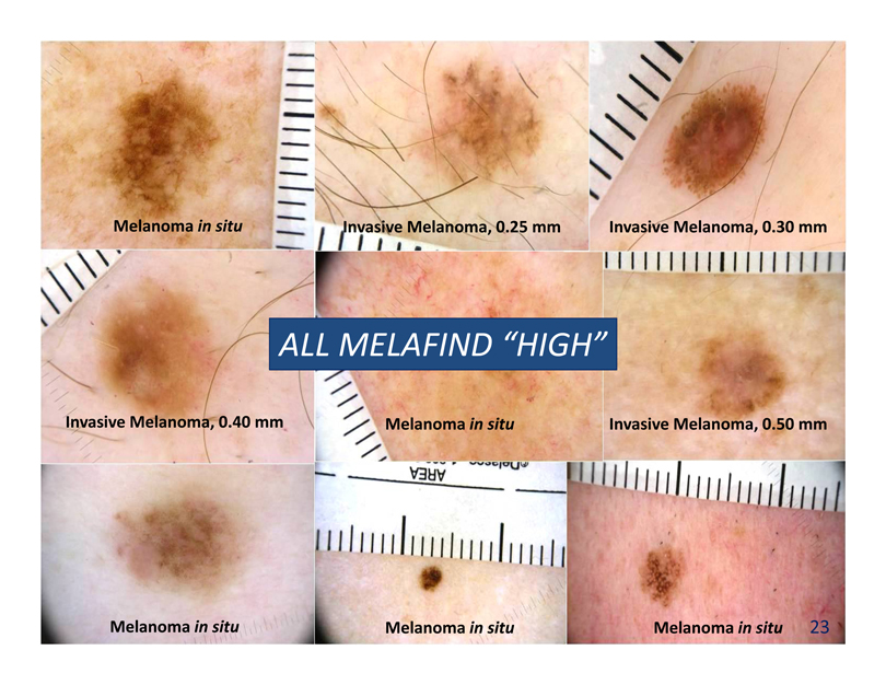
Melanoma in situ
Invasive Melanoma, 0.25 mm
Invasive Melanoma, 0.30 mm
Invasive Melanoma, 0.40 mm
ALL MELAFIND “HIGH”
Melanoma in situ
Invasive Melanoma, 0.50 mm
Melanoma in situ
Melanoma in situ
Melanoma in situ
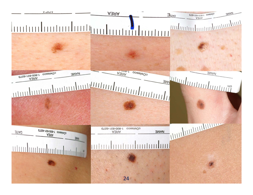
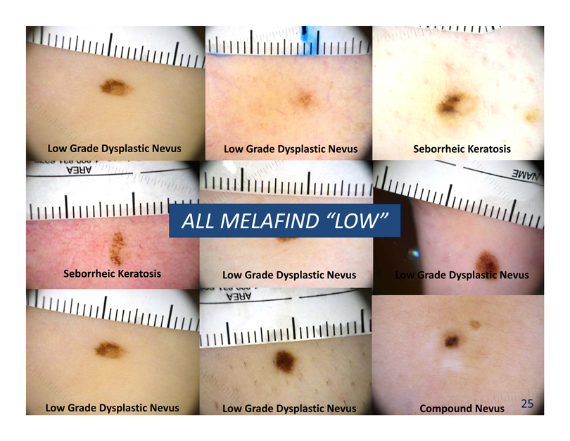
Low Grade Dysplastic Nevus
Low Grade Dysplastic Nevus
Seborrheic Keratosis
Seborrheic Keratosis
ALL MELAFIND “LOW”
Low Grade Dysplastic Nevus
Low Grade Dysplastic Nevus
Low Grade Dysplastic Nevus
Low Grade Dysplastic Nevus
Compound Nevus
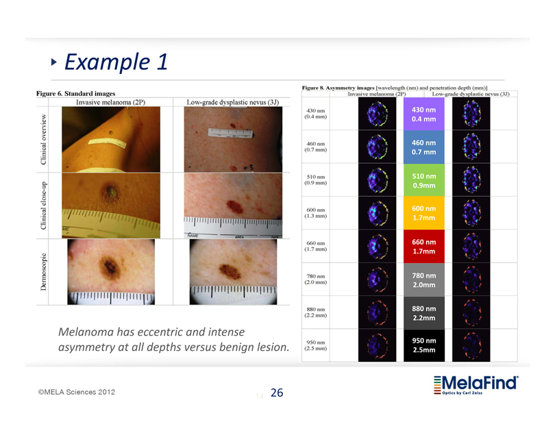
Example 1 Figure 6. Standard images Invasive melanoma (2P) Low-grade dysplastic nevus (3J) Dermoscopic Clinical close-up Clinical overview Melanoma has eccentric and intense asymmetry at all depths versus benign lesion. Figure 8. Asymmetry images [wavelength (mm) and penetration (mm) and penetraction depth (mm)] Invasive melanoma (2P) Low-grade dysplastic nevus (3J) 430 nm (0.4mm) 460 nm (0.7 mm) 510 nm (0.9 mm) 600 nm (1.3 mm) 660 nm (1.7 mm) 780 nm (2.0 mm) 880 nm (2.2 mn) 950 nm (2.5 mm) 430 nm (0.4mm) 460 nm (0.7 mm) 510 nm (0.9 mm) 600 nm (1.7 mm) 660 nm (1.7 mm) 780 nm (2.0 mm) 880 nm (2.2 mm) 950 nm (2.5 mm) © MELA Sciences 2012 MelaFind Optics by Carl Zelss 26
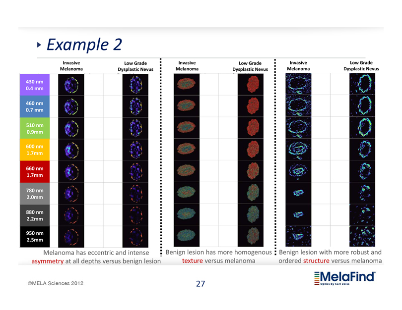
Example 2 (2.5 mm) 430 nm (0.4mm) 460 nm (0.7 mm) 510 nm (0.9 mm) 600 nm (1.7 mm) 660 nm (1.7 mm) 780 nm (2.0 mm) 880 nm (2.2 mn) 950 nm (2.5 mm) © MELA Sciences 2012 Invasive Melanoma Low Grade Dysplastic Nevus Invasive Melanoma Low Grade Dysplastic Nevus Invasive Melanoma Low Grade Dysplastic Nevus Melanoma has eccentric and intense asymmetry at all depths versus benign lesion Benign lesion has more homogenous texture versus melanoma Benign lesion with more robust and ordered structure versus melanoma MelaFind Optics by Carl Zelss
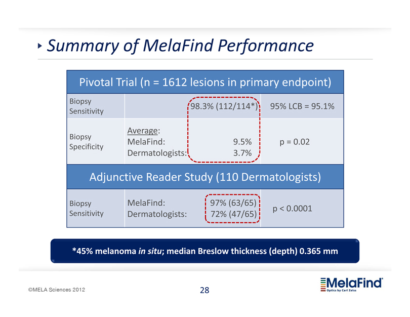
Summary of MelaFind Performance Pivotal Trial (n = 1612 lesions in primary endpoint) Biopsy 98.3% (112/114*) 95% LCB = 95.1% Sensitivity Average: Biopsy MelaFind: 9.5% p = 0.02 Specificity Dermatologists: 3.7% Adjunctive Reader Study (110 Dermatologists) Biopsy MelaFind: 97% (63/65) p < 0.0001 Sensitivity Dermatologists: 72% (47/65) *45% melanoma in situ; median Breslow thickness (depth) 0.365 mm 28 MelaFind optics by Carl Zeiss MELA Sciences 2012
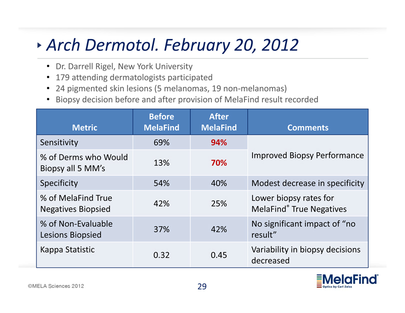
Arch Dermotol. February 20, 2012 Dr. Darrell Rigel, New York University 179 attending dermatologists participated 24 pigmented skin lesions (5 melanomas, 19 non-melanomas) Biopsy decision before and after provision of MelaFind result recorded Before After Metric MelaFind MelaFind Comments Sensitivity 69% 94% % of Derms who Would Improved Biopsy Performance 13% 70% Biopsy all 5 MM’s Specificity 54% 40% Modest decrease in specificity % of MelaFind True Lower biopsy rates for 42% 25% Negatives Biopsied MelaFind® True Negatives % of Non-Evaluable No significant impact of no 37% 42% Lesions Biopsied result” Kappa Statistic Variability in biopsy decisions 0.32 0.45 decreased 29 MelaFind optics by Carl Zeiss MELA Sciences 2012

Published in Premier Academic Journals STUDY Online First The Performance of MetaFind A Prospective Multicenter Study Gary Monheit, MD; Armand B. Cognetta, MD; Laura Ferris, MD, Mary Martini, MD; James M. Grichnik, MD, PhD; Martin Mihm, Roy King, MD; Alicia Toledano, ScD; Nikolai Kabelev, BCSc; Objective: To demonstrate the safety and effectiveness of Melafind, a noninvasive and objective computervision system designed to aid in detection of early pigmented cutaneous melanoma. Design: a prospective, multicenter, blinded study. The diagnostic performance of Mela find and of study clinicians was evaluated using the histologic reference standard. Standard images and patient information for a subset of 50 randomly selected lesions (25 melanomas) were used in a reader study of 39 independent deramatologists to estimate clinicians’ biopsy sensitivity to melanoma. Setting: Three academic and 4 community practices in the United States with expertise in management of pigmented skin lesions. Patients: A total of 1383 patients with 18311 lesions enrolled from January 2007 to July 2008; 1632 lesions (including 127 melanomas—45% in situ—with median Breslow thickness of invasive lesions, 0.36 mm) were eligible and evaluable for the study end points. ARCHIVES OF DERMATOLOGY Online First: February 20, 2012 Research Letters TABLE OF CONTENTS> ONLINE FIRST Impact of Guidance From a Computer-Aided Multispectral Digital Skin Lesion Analysis Device on Decision to Biopsy Lesions Clinically Suggestive of Melanoma Darrell S. Rigel, MD, MS; Mrinalini Roy, BA; Jane Yoo, MD MPP; Clay J. Cockerell, MD; June K. Robinson, MD; Richard White, MA Arch Dermatol. Published online February 20, 2012. Dol: 10.1001/archdermatol.2011.3388 A major challenge faced daily by clinical dermatologists is to determine which pigmented lesions are appropriate for biopsy. The present study was designed to determine the effect of guidance Inc)1 on dermatologists’ decision to biopsy a pigmented lesion and the impact of the imformation provided by the device on the associated melanoma blospy sensitivity and specificity. MelaFind uses light from visible to near-infrared wavelengths to image up to 2.5 mm beneath the skin and analyzes images from subbands of these wavelengths to provide information about the lesions’s level of structural disorder. The device provides an output of “positive” or “negative” as an additional piece of data that can be integrated into the blospy decision. © MELA Sciences 2012 MelaFind Optics by Carl zeiss
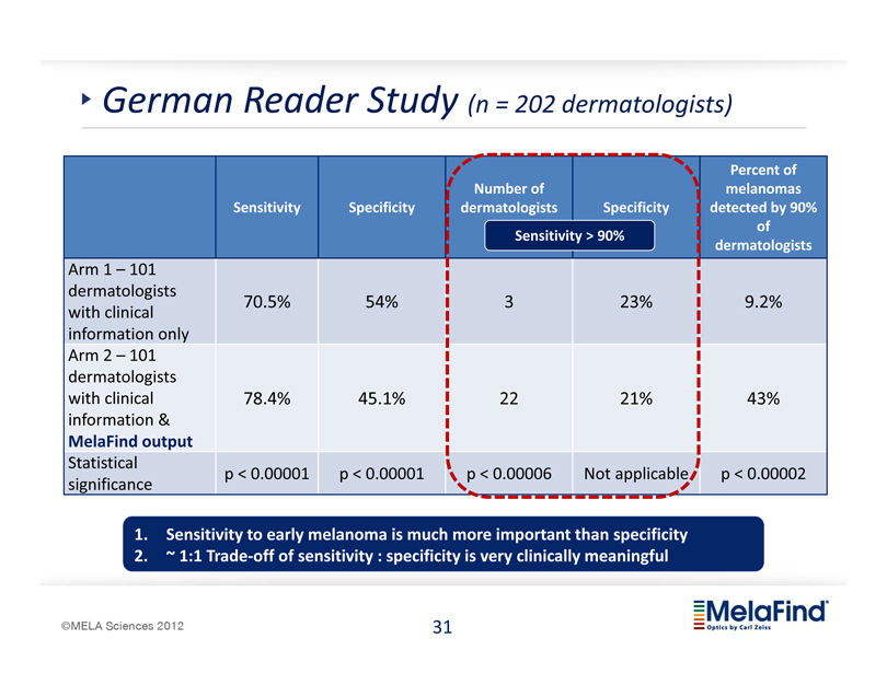
German Reader Study (n = 202 dermatologists) Percent of Number of melanomas Sensitivity Specificity dermatologists Specificity detected by 90% of Sensitivity > 90% dermatologists Arm 1 – 101 dermatologists 70.5% 54% 3 23% 9.2% with clinical information only Arm 2 – 101 dermatologists with clinical 78.4% 45.1% 22 21% 43% information & MelaFind output Statistical p < 0.00001 p < 0.00001 p < 0.00006 Not applicable p < 0.00002 significance 1. Sensitivity to early melanoma is much more important than specificity 2. ~ 1:1 Trade off of sensitivity : specificity is very clinically meaningful 31 MelaFind optics by Carl Zeiss
MELA Sciences 2012
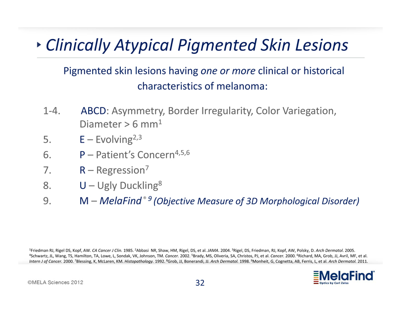
Clinically Atypical Pigmented Skin Lesions Pigmented skin lesions having one or more clinical or historical characteristics of melanoma: 14. ABCD: Asymmetry, Border Irregularity, Color Variegation, Diameter > 6 mm1 5. E – Evolving2,3 6. P – Patient’s Concern4,5,6 7. R – Regression7 8. U – Ugly Duckling8 9. M – MelaFind ® 9 (Objective Measure of 3D Morphological Disorder) 1Friedman RJ, Rigel DS, Kopf, AW. CA Cancer J Clin. 1985. 2Abbasi NR, Shaw, HM, Rigel, DS, et al. JAMA. 2004. 3Rigel, DS, Friedman, RJ, Kopf, AW, Polsky, D. Arch
Dermatol. 2005. 4Schwartz, JL, Wang, TS, Hamilton, TA, Lowe, L, Sondak, VK, Johnson, TM. Cancer. 2002. 5Brady, MS, Oliveria, SA, Christos, PJ, et al. Cancer. 2000. 6Richard, MA, Grob, JJ, Avril, MF, et al. Intern J of Cancer. 2000. 7Blessing, K, McLaren, KM. Histopathology. 1992. 8Grob, JJ, Bonerandi, JJ. Arch Dermatol. 1998. 9Monheit, G, Cognetta, AB, Ferris, L, et al. Arch Dermatol. 2011. 32 MelaFind optics by Carl Zeiss
MELA Sciences 2012
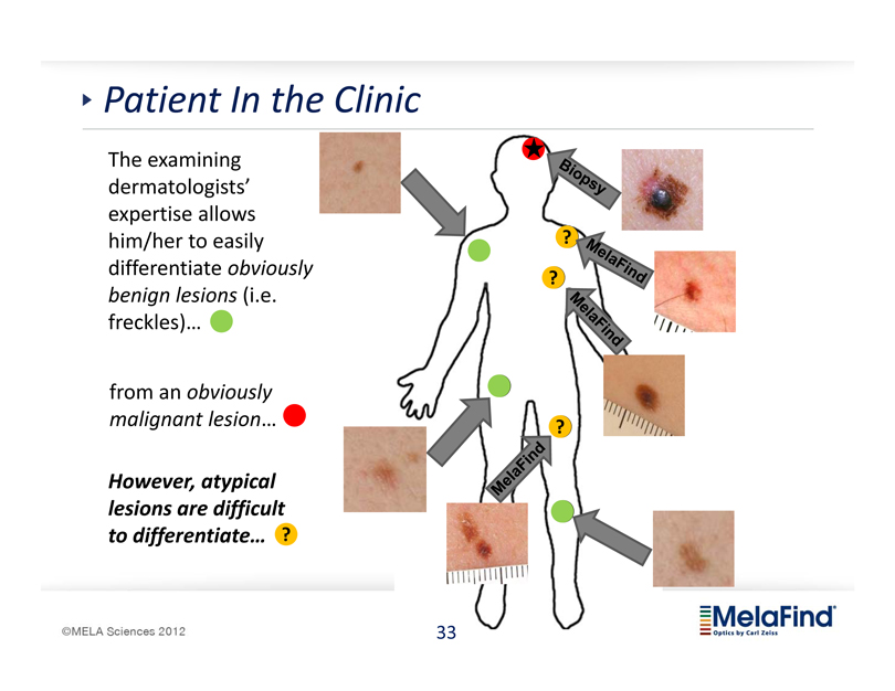
Patient In the Clinic The examining dermatologists’ expertise allows him/her to easily differentiate obviously benign lesions (i.e. freckles)… from an obviously malignant lesion… However, atypical lesions are difficult to differentiate… 33 MelaFind optics by Carl Zeiss
MELA Sciences 2012
Summary Summary Market for melanoma detection is large and growing Significant unmet medical need MelaFind® is a breakthrough product for early detection Largest positive prospective trial ever performed in melanoma detection Launched March 7, 2012 – US & Germany Goal: by March 31, 2013 – 200 systems in US and 75 Germany On target; medical utility in customer hands established Strong business model for commercialization Well capitalized 34 MelaFind optics by Carl Zeiss
MELA Sciences 2012
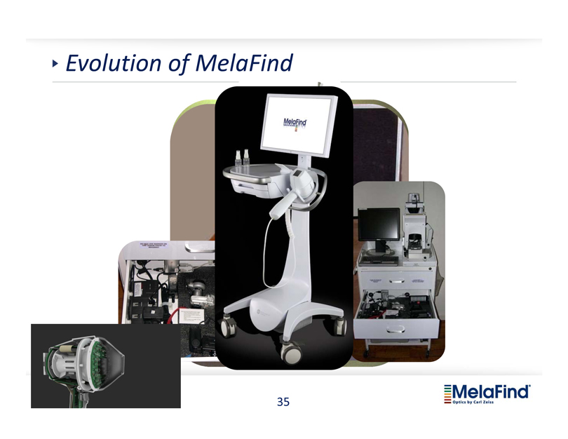
Evolution of MelaFind MelaFind optics by Carl Zeiss
MELA Sciences 2012