Exhibit 99.1
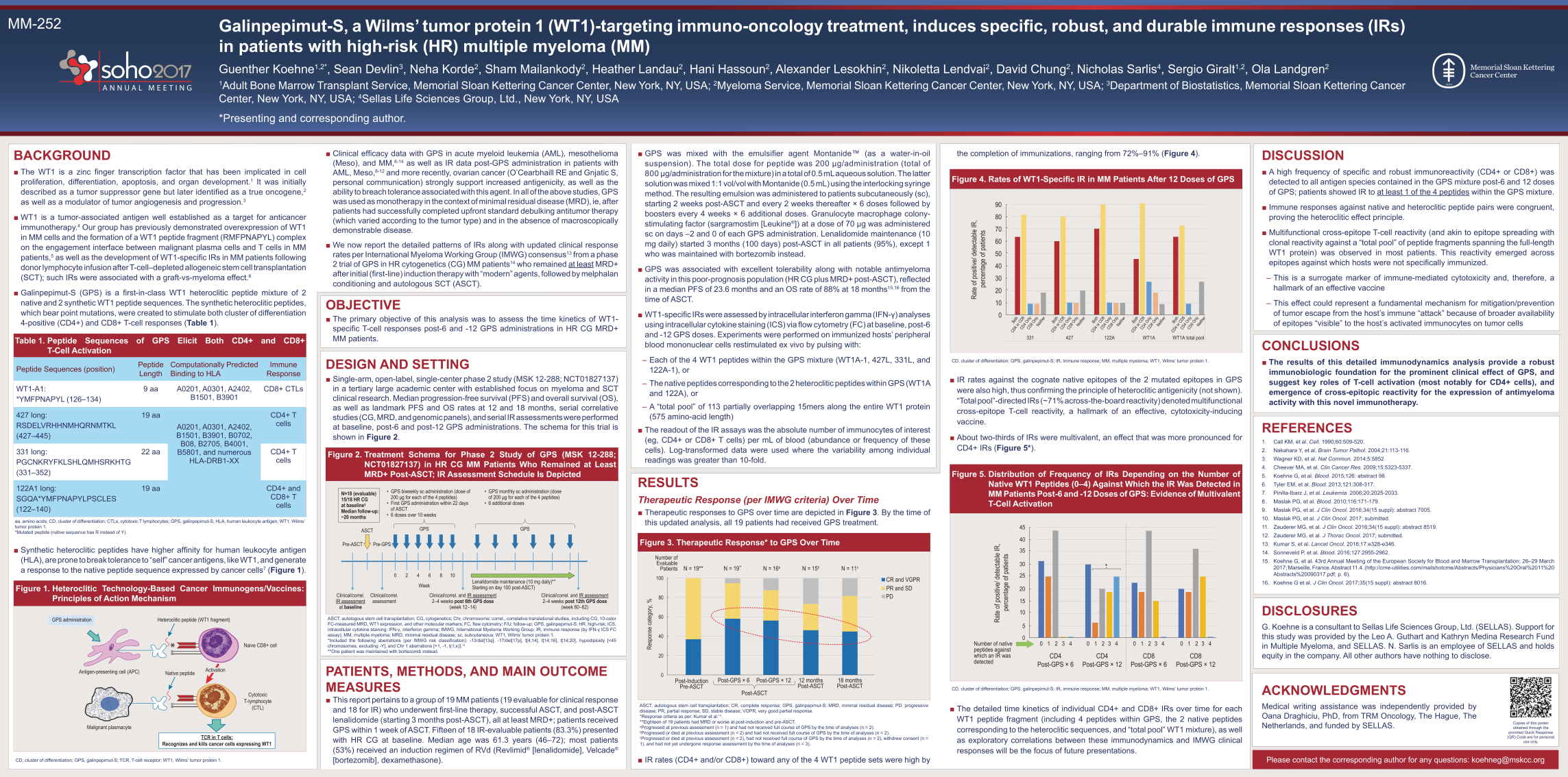
MM-252 Galinpepimut-S, a Wilms’ tumor protein 1 (WT1)-targeting immuno-oncology treatment, induces specific, robust, and durable immune responses (IRs) in patients with high-risk (HR) multiple myeloma (MM) Guenther Koehne1,2*, Sean Devlin3, Neha Korde2, Sham Mailankody2, Heather Landau2, Hani Hassoun2, Alexander Lesokhin2, Nikoletta Lendvai2, David Chung2, Nicholas Sarlis4, Sergio Giralt1,2, Ola Landgren2 1Adult Bone Marrow Transplant Service, Memorial Sloan Kettering Cancer Center, New York, NY, USA; 2Myeloma Service, Memorial Sloan Kettering Cancer Center, New York, NY, USA; 3Department of Biostatistics, Memorial Sloan Kettering Cancer Center, New York, NY, USA; 4Sellas Life Sciences Group, Ltd., New York, NY, USA *Presenting and corresponding author. BACKGROUND The WT1 is a zinc finger transcription factor that has been implicated in cell proliferation, differentiation, apoptosis, and organ development.1 It was initially described as a tumor suppressor gene but later identified as a true oncogene,2 as well as a modulator of tumor angiogenesis and progression.3 WT1 is a tumor-associated antigen well established as a target for anticancer immunotherapy.4 Our group has previously demonstrated overexpression of WT1 in MM cells and the formation of a WT1 peptide fragment (RMFPNAPYL) complex on the engagement interface between malignant plasma cells and T cells in MMpatients,5 as well as the development of WT1-specific IRs in MM patients following donor lymphocyte infusion after T-cell–depleted allogeneic stem cell transplantation (SCT); such IRs were associated with a graft-vs-myeloma effect.6 Galinpepimut-S (GPS) is a first-in-class WT1 heteroclitic peptide mixture of 2 native and 2 synthetic WT1 peptide sequences. The synthetic heteroclitic peptides, which bear point mutations, were created to stimulate both cluster of differentiation 4-positive (CD4+) and CD8+ T-cell responses (Table 1). Table 1. Peptide Sequences of GPS Elicit Both CD4+ and CD8+ T-Cell Activation Peptide Sequences (position) Peptide Length Computationally Predicted Binding to HLA Immune Response WT1-A1: *YMFPNAPYL (126–134) 9 aa A0201, A0301, A2402, B1501, B3901 CD8+ CTLs 427 long: RSDELVRHHNMHQRNMTKL (427–445) 19 aa A0201, A0301, A2402, B1501, B3901, B0702, B08, B2705, B4001, B5801, and numerous HLA-DRB1-XX CD4+ T cells 331 long: PGCNKRYFKLSHLQMHSRKHTG (331–352) 22 aa CD4+ T cells 122A1 long: SGQA*YMFPNAPYLPSCLES (122–140) 19 aa CD4+ and CD8+ T cells aa, amino acids; CD, cluster of differentiation; CTLs, cytotoxic T lymphocytes; GPS, galinpepimut-S; HLA, human leukocyte antigen; WT1, Wilms’ tumor protein 1. *Mutated peptide (native sequence has R instead of Y). Synthetic heteroclitic peptides have higher affinity for human leukocyte antigen (HLA), are prone to break tolerance to “self” cancer antigens, like WT1, and generate a response to the native peptide sequence expressed by cancer cells7 (Figure 1) Figure 1. Heteroclitic Technology-Based Cancer Immunogens/Vaccines: Principles of Action Mechanism Heteroclitic peptide (WT1 fragment) Native peptide Naive CD8+ cell Cytotoxic T-lymphocyte (CTL) Malignant plasmacyte Antigen-presenting cell (APC) GPS administration TCR in T cells: Recognizes and kills cancer cells expressing WT1 Activation CD, cluster of differentiation; GPS, galinpepimut-S; TCR, T-cell receptor; WT1, Wilms’ tumor protein 1. Clinical efficacy data with GPS in acute myeloid leukemia (AML), mesothelioma (Meso), and MM,8-14 as well as IR data post-GPS administration in patients with AML, Meso,8-12 and more recently, ovarian cancer (O’Cearbhaill RE and Gnjatic S, personal communication) strongly support increased antigenicity, as well as the ability to breach tolerance associated with this agent. In all of the above studies, GPS was used as monotherapy in the context of minimal residual disease (MRD), ie, after patients had successfully completed upfront standard debulking antitumor therapy (which varied according to the tumor type) and in the absence of macroscopically demonstrable disease. We now report the detailed patterns of IRs along with updated clinical response rates per International Myeloma Working Group (IMWG) consensus13 from a phase 2 trial of GPS in HR cytogenetics (CG) MM patients14 who remained at least MRD+ after initial (first-line) induction therapy with “modern” agents, followed by melphalan conditioning and autologous SCT (ASCT). OBJECTIVE The primary objective of this analysis was to assess the time kinetics of WT1- specific T-cell responses post-6 and -12 GPS administrations in HR CG MRD+ MM patients. DESIGN AND SETTING Single-arm, open-label, single-center phase 2 study (MSK 12-288; NCT01827137) in a tertiary large academic center with established focus on myeloma and SCT clinical research. Median progression-free survival (PFS) and overall survival (OS), as well as landmark PFS and OS rates at 12 and 18 months, serial correlative studies (CG, MRD, and genomic panels), and serial IR assessments were performed at baseline, post-6 and post-12 GPS administrations. The schema for this trial is shown in Figure 2. Figure 2. Treatment Schema for Phase 2 Study of GPS (MSK 12-288; NCT01827137) in HR CG MM Patients Who Remained at Least MRD+ Post-ASCT; IR Assessment Schedule Is Depicted ASCT Pre-ASCT Pre-GPS 0 2 4 6 8 10 GPS Week Clinical/correl. assessment Clinical/correl. and IR assessment 2–4 weeks post 6th GPS dose (week 12–14) Clinical/correl. and IR assessment 2–4 weeks post 12th GPS dose (week 60–62) Lenalidomide maintenance (10 mg daily)** Starting on day 100 post-ASCT) Clinical/correl. IR assessment at baseline N=18 (evaluable) 15/18 HR CG at baseline Median follow-up: ~20 months GPS GPS biweekly sc administration (dose of 200 g for each of the 4 peptides) First GPS administration within 22 days of ASCT 6 doses over 10 weeks GPS monthly sc administration (dose of 200 g for each of the 4 peptides) 6 additional doses ASCT, autologous stem cell transplantation; CG, cytogenetics; Chr, chromosome; correl., correlative translational studies, including CG, 10-color FC-measured MRD, WT1 expression, and other molecular markers; FC, flow cytometry; F/U: follow-up; GPS, galinpepimut-S; HR, high-risk; ICS, intracellular cytokine staining; IFN-, interferon gamma; IMWG, International Myeloma Working Group; IR, immune response (by IFN- ICS FC assay); MM, multiple myeloma; MRD, minimal residual disease; sc, subcutaneous; WT1, Wilms’ tumor protein 1. *Included the following aberrations (per IMWG risk classification): -13/del[13q], -17/del[17p], t[4;14], t[14;16], t[14;20], hypodiploidy [<45 chromosomes, excluding -Y], and Chr 1 aberrations [+1, -1, t(1;x)].14 **One patient was maintained with bortezomib instead. PATIENTS, METHODS, AND MAIN OUTCOME MEASURES This report pertains to a group of 19 MM patients (19 evaluable for clinical response and 18 for IR) who underwent first-line therapy, successful ASCT, and post-ASCT lenalidomide (starting 3 months post-ASCT), all at least MRD+; patients received GPS within 1 week of ASCT. Fifteen of 18 IR-evaluable patients (83.3%) presented with HR CG at baseline. Median age was 61.3 years (46–72); most patients (53%) received an induction regimen of RVd (Revlimid® [lenalidomide], Velcade® [bortezomib], dexamethasone). GPS was mixed with the emulsifier agent MontanideTM (as a water-in-oil suspension). The total dose for peptide was 200 g/administration (total of 800 g/administration for the mixture) in a total of 0.5 mL aqueous solution. The latter solution was mixed 1:1 vol/vol with Montanide (0.5 mL) using the interlocking syringe method. The resulting emulsion was administered to patients subcutaneously (sc), starting 2 weeks post-ASCT and every 2 weeks thereafter × 6 doses followed by boosters every 4 weeks × 6 additional doses. Granulocyte macrophage colonystimulating factor (sargramostim [Leukine®]) at a dose of 70 g was administered sc on days –2 and 0 of each GPS administration. Lenalidomide maintenance (10 mg daily) started 3 months (100 days) post-ASCT in all patients (95%), except 1 who was maintained with bortezomib instead. GPS was associated with excellent tolerability along with notable antimyeloma activity in this poor-prognosis population (HR CG plus MRD+ post-ASCT), reflected in a median PFS of 23.6 months and an OS rate of 88% at 18 months15,16 from the time of ASCT. WT1-specific IRs were assessed by intracellular interferon gamma (IFN-) analyses using intracellular cytokine staining (ICS) via flow cytometry (FC) at baseline, post-6 and -12 GPS doses. Experiments were performed on immunized hosts’ peripheral blood mononuclear cells restimulated ex vivo by pulsing with: ––Each of the 4 WT1 peptides within the GPS mixture (WT1A-1, 427L, 331L, and 122A-1), or ––The native peptides corresponding to the 2 heteroclitic peptides within GPS (WT1A and 122A), or –A “total pool” of 113 partially overlapping 15mers along the entire WT1 protein (575 amino-acid length) The readout of the IR assays was the absolute number of immunocytes of interest (eg, CD4+ or CD8+ T cells) per mL of blood (abundance or frequency of these cells). Log-transformed data were used where the variability among individual readings was greater than 10-fold. RESULTS Therapeutic Response (per IMWG criteria) Over TimeTherapeutic responses to GPS over time are depicted in Figure 3. By the time of this updated analysis, all 19 patients had received GPS treatment. Figure 3. Therapeutic Response* to GPS Over Time 100 80 60 40 20 Response category, % 0 Post-Induction Pre-ASCT Number of Evaluable Patients Post-GPS × 12 Post-ASCT Post-GPS × 6 12 months Post-ASCT 18 months Post-ASCT N = 19** N = 19** N = 16a N = 15b N = 11c CR and VGPR PR and SD PD
ASCT, autologous stem cell transplantation; CR, complete response; GPS, galinpepimut-S; MRD, minimal residual disease; PD, progressive disease; PR, partial response; SD, stable disease; VGPR, very good partial response. *Response criteria as per: Kumar et al.13. **Eighteen of 19 patients had MRD or worse at post-induction and pre-ASCT. a Progressed at previous assessment (n = 1) and had not received full course of GPS by the time of analyses (n = 2). bProgressed or died at previous assessment (n = 2) and had not received full course of GPS by the time of analyses (n = 2). cProgressed or died at previous assessment (n = 2), had not received full course of GPS by the time of analyses (n = 2), withdrew consent (n = 1), and had not yet undergone response assessment by the time of analyses (n = 3). IR rates (CD4+ and/or CD8+) toward any of the 4 WT1 peptide sets were high by the completion of immunizations, ranging from 72%–91% (Figure 4). Figure 4. Rates of WT1-Specific IR in MM Patients After 12 Doses of GPS 90 40 50 60 70 80 30 20 10 0 Rate of positive/ detectable IR, percentage of patients Both CD4 or CD8 CD4 Only CD8 Only Neither Both CD4 or CD8 CD4 Only CD8 Only Neither Both CD4 or CD8 CD4 Only CD8 Only Neither Both CD4 or CD8 CD4 Only CD8 Only Neither Both CD4 or CD8 CD4 Only CD8 Only Neither 331 427 122A WT1A WT1A total pool CD, cluster of differentiation; GPS, galinpepimut-S; IR, immune response; MM, multiple myeloma; WT1, Wilms’ tumor protein 1. IR rates against the cognate native epitopes of the 2 mutated epitopes in GPS were also high, thus confirming the principle of heteroclitic antigenicity (not shown). “Total pool”-directed IRs (~71% across-the-board reactivity) denoted multifunctional cross-epitope T-cell reactivity, a hallmark of an effective, cytotoxicity-inducing vaccine. About two-thirds of IRs were multivalent, an effect that was more pronounced for CD4+ IRs (Figure 5*). Figure 5. Distribution of Frequency of IRs Depending on the Number of Native WT1 Peptides (0–4) Against Which the IR Was Detected in MM Patients Post-6 and -12 Doses of GPS: Evidence of Multivalent T-Cell Activation 45 20 25 30 35 40 15 10 5 0 0 1 2 CD4 Post-GPS × 6 CD4 Post-GPS × 12 CD8 Post-GPS × 6 CD8 Post-GPS × 12 3 4 0 1 2 3 4 0 1 2 3 4 0 1 2 3 4 * Number of native peptides against which an IR was detected Rate of positive/ detectable IR, percentage of patients CD, cluster of differentiation; GPS, galinpepimut-S; IR, immune response; MM, multiple myeloma; WT1, Wilms’ tumor protein 1. The detailed time kinetics of individual CD4+ and CD8+ IRs over time for each WT1 peptide fragment (including 4 peptides within GPS, the 2 native peptides corresponding to the heteroclitic sequences, and “total pool” WT1 mixture), as well as exploratory correlations between these immunodynamics and IMWG clinical responses will be the focus of future presentations. DISCUSSION A high frequency of specific and robust immunoreactivity (CD4+ or CD8+) was detected to all antigen species contained in the GPS mixture post-6 and 12 doses of GPS; patients showed IR to at least 1 of the 4 peptides within the GPS mixture. Immune responses against native and heteroclitic peptide pairs were congruent, proving the heteroclitic effect principle. Multifunctional cross-epitope T-cell reactivity (and akin to epitope spreading with clonal reactivity against a “total pool” of peptide fragments spanning the full-length WT1 protein) was observed in most patients. This reactivity emerged across epitopes against which hosts were not specifically immunized. ––This is a surrogate marker of immune-mediated cytotoxicity and, therefore, a hallmark of an effective vaccine ––This effect could represent a fundamental mechanism for mitigation/prevention of tumor escape from the host’s immune “attack” because of broader availability of epitopes “visible” to the host’s activated immunocytes on tumor cells CONCLUSIONS The results of this detailed immunodynamics analysis provide a robust immunobiologic foundation for the prominent clinical effect of GPS, and suggest key roles of T-cell activation (most notably for CD4+ cells), and emergence of cross-epitopic reactivity for the expression of antimyeloma activity with this novel immunotherapy. REFERENCES 1. Call KM, et al. Cell. 1990;60:509-520. 2. Nakahara Y, et al. Brain Tumor Pathol. 2004;21:113-116. 3. Wagner KD, et al. Nat Commun. 2014;5:5852. 4. Cheever MA, et al. Clin Cancer Res. 2009;15:5323-5337. 5. Koehne G, et al. Blood. 2015;126: abstract 98. 6. Tyler EM, et al. Blood. 2013;121:308-317. 7. Pinilla-Ibarz J, et al. Leukemia. 2006;20:2025-2033. 8. Maslak PG, et al. Blood. 2010;116:171-179. 9. Maslak PG, et al. J Clin Oncol. 2016;34(15 suppl): abstract 7005. 10. Maslak PG, et al. J Clin Oncol. 2017; submitted. 11. Zauderer MG, et al. J Clin Oncol. 2016;34(15 suppl): abstract 8519. 12. Zauderer MG, et al. J Thorac Oncol. 2017; submitted. 13. Kumar S, et al. Lancet Oncol. 2016;17:e328-e346. 14. Sonneveld P, et al. Blood. 2016;127:2955-2962. 15. Koehne G, et al. 43rd Annual Meeting of the European Society for Blood and Marrow Transplantation; 26–29 March 2017; Marseille, France. Abstract 11.4. (http://cme-utilities.com/mailshotcme/Abstracts/Physicians%20Oral%2011%20 Abstracts%20090317.pdf; p. 6). 16. Koehne G et al. J Clin Oncol. 2017;35(15 suppl): abstract 8016. DISCLOSURES G. Koehne is a consultant to Sellas Life Sciences Group, Ltd. (SELLAS). Support for this study was provided by the Leo A. Guthart and Kathryn Medina Research Fund in Multiple Myeloma, and SELLAS. N. Sarlis is an employee of SELLAS and holds equity in the company. All other authors have nothing to disclose. ACKNOWLEDGMENTS Medical writing assistance was independently provided by Oana Draghiciu, PhD, from TRM Oncology, The Hague, The Netherlands, and funded by SELLAS. Please contact the corresponding author for any questions: koehneg@mskcc.org Copies of this poster obtained through the provided Quick Response (QR) Code are for personal use only.
Galinpepimut-S, a WT1-targeting immuno-Oncology Treatment, Induces Specific, Robust and Durable Immune Responses (IRs) in Patients (Pts) With High-Risk (HR) Multiple Myeloma (MM)
Guenther Koehne1, 2, Sean Devlin3, Neha Korde2, Sham Mailankody2, Heather Landau2, Hani Hassoun4, Alexander Lesokhin2, Nikoletta Lendvai2, David Chung2, Nicholas Sarlis5, Sergio Giralt6, Ola Landgren2.
1Cytotherapy Laboratory, Memorial Sloan Kettering Cancer Center (MSKCC), New York, NY, USA,2Myeloma Service, Memorial Sloan Kettering Cancer Center (MSKCC), New York, NY, USA,3Department of Biostatistics, Memorial Sloan Kettering Cancer Center (MSKCC), New York, NY, USA,4Lymphoma Service, Memorial Sloan Kettering Cancer Center (MSKCC), New York, NY, USA,5Sellas Life Sciences Group, Ltd., New York, NY, USA,6Adult Bone Marrow Transplant Service, Memorial Sloan Kettering Cancer Center (MSKCC), New York, NY, USA.
Presenter: undelined
ABSTRACT SUBMITTED TO THE SOHO 2017 MEETING, Houston, TX, Sep. 13-16, 2017
https://soho2017.com/
KEY WORDS: WT1, peptide, high risk, immunotherapy, immune response.
CONTEXT: MM pts with HR cytogenetics (CG) also harboring minimal residual disease (MRD+) after upfront Rx followed by (f/b) ASCT continue to experience poor clinical outcomes, despite the introduction of IMiD maintenance. We have targeted a major MM antigenic target, Wilms Tumor-1 protein (WT1) with a novel immunotherapy.
OBJECTIVE: MM pts immunized with the WT1 heteroclitic peptide mixture galinpepimut-S (GPS) post-ASCT showed an 88% OS at 18 months (mo) and median PFS of 23.6 mo (EBMT & ASCO, 2017). We now report the detailed patterns of corresponding IRs.
DESIGN: Open-label, single-center Phase 2 study. PFS, OS, serial CG, and MRD & IR assessments followed up for a median of 18 mo.
SETTING: Tertiary large oncology center.
PATIENTS OR OTHER PARTICIPANTS: 18 MM pts post-ASCT f/b lenalidomide starting 3 months (mo) post-ASCT (all at least MRD+). 15/18 pts presented with HR-CG.
INTERVENTIONS: GPS was administered with montanide s.c. starting 2 wks post-ASCT and q2 wks thereafter x 6 doses f/b boosters q4 wks x 6 additional doses. GM-CSF was also given. GPS consisted of 4 peptides: WT1A-1(*); 427L (long); 331L, and 122A1L(*). Two of the 4 peptides were mutated (heteroclitic; *) to induce stronger HLA-binding/ reduce tolerance.
MAIN OUTCOMES MEASURES:WT1-specific IRs were assessed by intracellular IFN-g analyses (baseline, post-6 & -12 GPS doses), using PBMC’s pulsed with: each of the 4 WT1 peptides in GPS; or the native peptides corresponding to the 2 heteroclitic ones; or a ‘total pool’ of overlapping 15mers along the entire WT1 protein.
RESULTS: IR rates (CD4 and/or CD8) toward any of the 3 WT1 peptide sets were high by the completion of immunizations, ranging from 72-91%; 2/3’s of IRs were multivalent. IR rates against the cognate native epitopes of the 2 mutated ones in GPS were also high, thus confirming the principle of heteroclitic antigenicity. ‘Total pool’-directed IRs denoted multifunctional cross-epitope T-cell reactivity, a hallmark of an effective, cytotoxicity-inducing vaccine.
CONCLUSIONS: The results of this detailed immunodynamics analysis provide a robust immunobiological foundation for the striking clinical effect of GPS, and suggest key roles of CD4 activation and emergence of cross-epitopic reactivity for antimyeloma activity with this therapy.
Support for this study was provided by Leo A. Guthart and Kathryn Medina Research Fund in Multiple Myeloma, and Sellas Life Sciences Group, Ltd.