EXHIBIT 99.2

“Evolving Role and Clinical Impact of ProstaScint®”
Michael J. Manyak, MD, FACS
Vice President, Medical Affairs
CYTOGEN Corp
ProstaScint®
Early development
Positive predictive value modest
Findings difficult to interpret without training
Significance of midabdominal signal
Importance of negative predictive value not appreciated
Limitations due to imaging technology, not the antibody
Slide # 2
Radioimmunoscintigraphy Advances
Significant improvements in resolution
Image co-registration
Outcomes data
Image-guided therapy
Slide # 3
Models for Prediction
PSA doubling time may suggest metastasis but does not give location or extent of disease
Nomogram prediction of lymph node metastasis based on path samples from very limited lymph node areas
Slide # 4
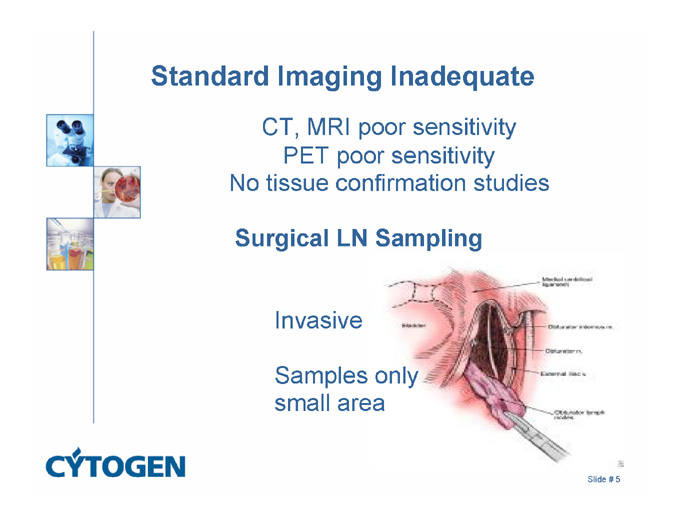
Standard Imaging Inadequate
CT, MRI poor sensitivity
PET poor sensitivity
No tissue confirmation studies
Surgical LN Sampling
Invasive
Samples only
small area
Slide # 5
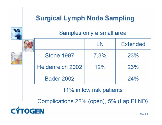
Surgical Lymph Node Sampling
Samples only a small area
LN
Extended
Stone 1997
7.3%
23%
Heidenreich 2002
12%
26%
Bader 2002
24%
11% in low risk patients
Complications 22% (open), 5% (Lap PLND)
Slide # 6
Surgery
Extended LN dissection still only samples selected areas
4.5% of LN in APR for colorectal cancer were actually prostate cancer (Murray, Am J Surg Path, 2004)
Extended LN dissection misses disease
43% 5 yr progression free rate (Allaf, J Urol 2004)
39% 4 yr progression free rate rate (Bader, J Urol 2003)
Modification: Radio-guided sentinel LN biopsy
Preliminary, PPV low, 15% False (-) breast accepted
(Weckermann, E Urol, 2005; Jeschke, J Urol, 2005)
Slide # 7
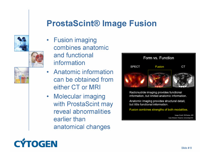
ProstaScint® Image Fusion
Fusion imaging combines anatomic and functional information
Anatomic information can be obtained from either CT or MRI
Molecular imaging with ProstaScint may reveal abnormalities earlier than anatomical changes
Slide # 8
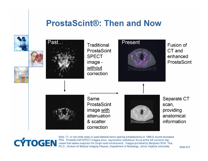
ProstaScint®: Then and Now
Present
Past…
Traditional ProstaScintSPECT image - without correction
Fusion of CT and enhanced ProstaScint
Separate CT scan, providing anatomical information
Same ProstaScint image with attenuation & scatter correction
Note: 71- yr old white male w/ post bilateral nerve sparing prostatectomy in 1996 & recent increased PSA. ProstaScint® SPECT images show asymmetric radiotracer focus at the left common iliac vessel that raises suspicion for lymph node involvement. Images provided by Benjamin M.W. Tsui, Ph.D., Division of Medical Imaging Physics, Department of Radiology, Johns Hopkins University
Slide # 9
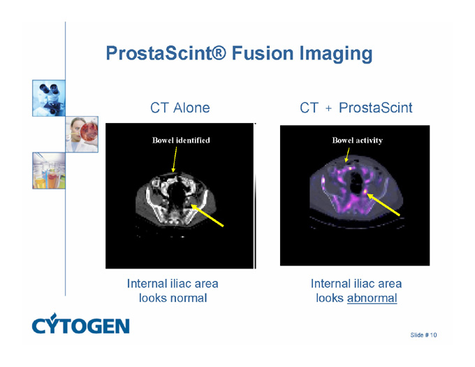
ProstaScint® Fusion Imaging
CT + ProstaScint
CT Alone
Bowel identified
Bowel activity
Internal iliac area looks normal
Internal iliac area looks abnormal
Slide # 10
Value of Image Co-Registration
Schettino et al., AJR 2004
58 pts, 74 of 161 + sites actually (–)
25 pts thought to be + were really (–) with fusion
Wong et al., AJR 2005
Image quality improved and interpretation simplified with fusion images
Better antibody localization using fusion images
Sodee et al., Clin Prostate Cancer 2005
Technique and experience with fusion images
49 pts, 83% accuracy with surgical confirmation
Localization accuracy doubled with fusion images
Slide # 11
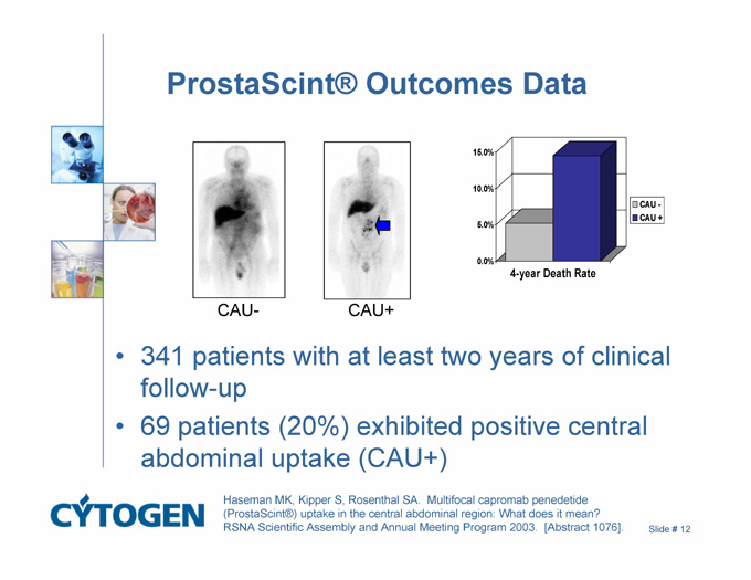
ProstaScint® Outcomes Data
CAU-
CAU+
15.0%
10.0%
5.0%
0.0%
CAU-
CAU+
4-year Death Rate
341 patients with at least two years of clinical follow-up
69 patients (20%) exhibited positive central abdominal uptake (CAU+)
Haseman MK, Kipper S, Rosenthal SA. Multifocal capromab penedetide (ProstaScint®) uptake in the central abdominal region: What does it mean? RSNA Scientific Assembly and Annual Meeting Program 2003. [Abstract 1076].
Slide # 12
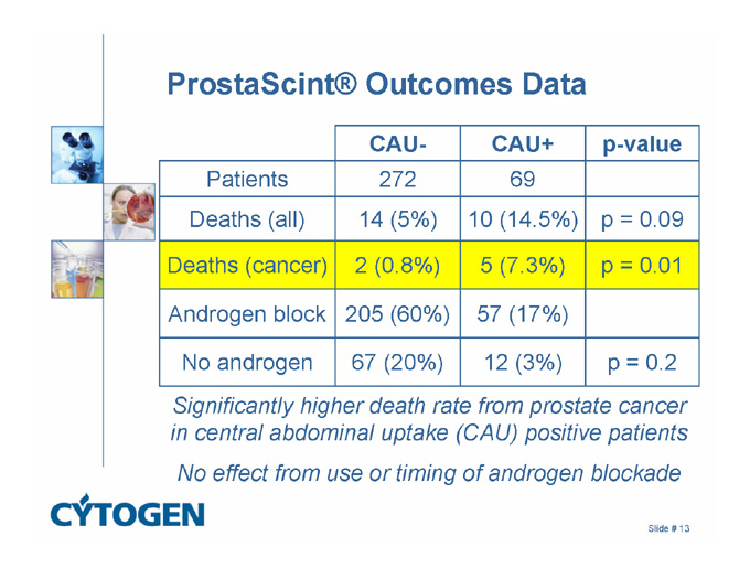
ProstaScint® Outcomes Data
CAU-
CAU+
p-value
Patients
272
69
Deaths (all)
14 (5%)
10 (14.5%)
p = 0.09
Deaths (cancer)
2 (0.8%)
5 (7.3%)
p = 0.01
Androgen block
205 (60%)
57 (17%)
No androgen
67 (20%)
12 (3%)
p = 0.2
Significantly higher death rate from prostate cancer in central abdominal uptake (CAU) positive patients
No effect from use or timing of androgen blockade
Slide # 13
Advantage to use of Fused Images
Jani et al. J Nuc Med 2004
107 pts, 3 yr biochemical free survival 85% versus 71%
Only fused scans used for treatment plan significant (p=0.04)
Schild et al. RSNA abstract 2004
43 pts, coregistration to increase or decrease dose
Low toxicity, preliminary PSA data
Slide # 14
Imaging: Clinical Outcomes
PSMA overexpression: Twice the occurrence and rate of biochemical recurrence (Ross, Clin CA Res, 2003)
Fused images: 83% sensitivity, tissue confirmed, doubled localization accuracy (Sodee,Clin Prost CA 2005)
Central abdominal signal: 3 fold increase in death (Haseman, 2005)
Fused image-guided IMRT (Schild, 2005)
Fused image-guided brachytherapy: 7 yr data (Ellis, 2005)
Slide # 15